| Normal Cardiac Long Axis View |
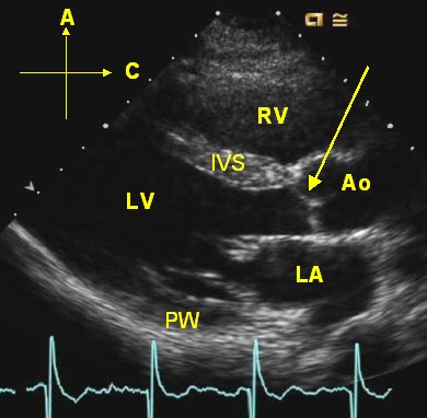 |
| The labeled still image ("Normal Long Axis") is a still frame
from an echocardiographic long axis view, a tomographic slice from the left ventricular apex to the base of the heart.
The labels represent: IVS - interventricular septum PW - posterior wall of the left ventricle Now open the video clip labeled "Normal AV" to see normal aortic valve motion. Open and compare that clip to the "Stenotic AV" clip. Notice the thickening of the valve leaflets and the limited valve excursion. This is a bicuspid aortic valve in a 40 year old man with mild aortic regurgitation and severe aortic stenosis. |